EOTRH: Equine Odontoclastic Tooth Resorption and Hypercementosis
By: Dr. Leah Limone, Northeast Equine Veterinary Dental Services, Topsfield, MA with Dr. Lydia Gray
What is EOTRH?
EOTRH is a syndrome in older horses resulting in disease and destruction of incisors and canine teeth, and sometimes cheek teeth. In order to better understand this recently recognized dental problem in senior horses, it may be helpful to define the terms used in its official name:
- Odontoclastic – refers to the cells (odontoclasts) that are involved in tooth resorption
- Resorption – the body’s internal process of destruction, disappearance, or dissolving of tissue
- Cementum – the bone-like substance covering the entire surface of the equine tooth, protecting it and helping to anchor it to the jawbone below the gumline
- Hypercementosis – abnormal, excessive formation of cementum
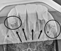
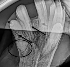
As the disease progresses, the roots of multiple teeth begin to resorb (dissolve), and the body tries to stabilize these teeth by laying down extra cementum. This results in hypercementosis, or bulbous swellings around the roots of affected teeth. These teeth become infected, abscess, and may loosen or even fracture. In severe cases there may also be significant extension of disease to the bone around the affected teeth. EOTRH is a slowly progressive disease, and it is unknown at this point what the trigger is that starts the process. It is believed that the process actually begins well before owners and veterinarians first start to recognize signs.
EOTRH is a very painful condition for affected horses, and chronically affected horses have weight loss, lack of appetite, attitude changes, hypersalivation, head shaking, discomfort in the bridle, and other signs. It is known that both periodontal disease and dental abscesses are painful, and this syndrome causes a significant degree of both, with usually multiple teeth affected. Because horses are quite stoic and adapt to this chronic pain, it is typically not until the pain is gone (when the affected teeth are extracted) that it is realized how much it was affecting them.
Diagnosis
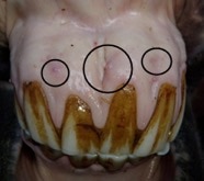
EOTRH is typically diagnosed on a routine veterinary dental exam. It is mostly identified in older horses (15+), but that is really because that is when the signs are more obvious. The veterinarians focused on dentistry that are looking closely for more subtle changes are diagnosing early cases in younger horses. Clinical signs include:
- hyperemia of the gums (reddening due to inflammation)
- petechia of the gums (small red dots) above the incisors
- oral ulcers of the gums, also above the incisors
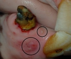
- drainage tracts (infection or abscess chronically draining)
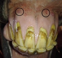
- receding gumlines (teeth look longer than they should normally, or they all look uneven lengths)
- deep pockets in the gumline (areas where the tooth has lost attachment to the bone)
- enlarged and inflamed tissue around the top of the teeth
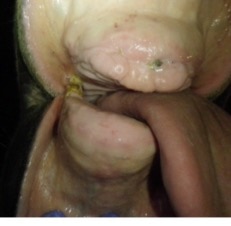
- bulbous, discolored, and/or cauliflower-like incisors
- fractured and/or loose teeth
- teeth with resorptive lesions visible (holes in the teeth, usually filled with feed)
- pus draining from areas of the gums or from teeth
- foul odor, bad breath
- sensitivity or discomfort identified on oral exam (teeth chattering, resistance to speculum pressure on incisors)
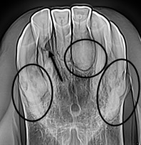
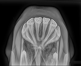
Radiographs are essential to making a diagnosis, because most of this process occurs below the gumline and cannot be seen without proper radiographic evaluation. Often there are more severe changes present radiographically than what are expected by looking at the teeth and gums, so there may be subtle changes that tip veterinarians off to see if there is something more going on. Radiographs will show whether the horse has more severe resorption or abscessation, or if there is significant hypercementosis present that could make extractions more difficult. Veterinarians use these images to then make a plan for extractions, whether they are staged if there are only a few teeth affected, or if all of the incisors and canines require extraction due to advanced disease. Once EOTRH is diagnosed and an extraction plan formulated, your veterinarian may also recommend baseline bloodwork including a test for Cushing’s Disease to be sure there are no underlying issues that may complicate healing.
Treatment
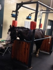
Mild cases of EOTRH or those in the early stages of the syndrome may simply be monitored with regular oral exams and radiographs as needed to follow the progression of the disease. However, staged or complete tooth extraction is recommended for moderate to severe cases. Regarding extractions, horses tolerate these very well. In most situations, horses are transported to a local veterinary clinic, teaching hospital, or surgical facility, where they are heavily sedated but remain standing in a set of stocks for safety and support. In addition, they are given plenty of local anesthesia so they cannot feel anything happening in the mouth.
Following these extractions, radiographs may be repeated to ensure that no diseased tissue is left behind and that remaining structures are healthy and intact. Pain medication and antibiotics are usually prescribed. These horses are typically eating a soft, soaked pelleted mash or chopped or pelleted hay before going home the same day, and back to normal feed the following day. These horses generally start feeling better within 24 hours of having chronically infected and painful teeth removed, and owners frequently report that there is a noticeable difference.
Aftercare Management and Diet
There is typically no diet change after extractions, as horses use their cheek teeth and not their incisors to chew hay or feed. They are still very much able to graze fresh grass as well as pick up hay and grain using their lips and tongue. However, as many of these cases involve older horses, regular annual or semiannual oral exams with a veterinarian should be continued to monitor and maintain the health of the rest of their cheek teeth. Horses that have undergone tooth extractions are typically completely healed between 4-6 weeks – with suture removal at around 10 days to 2 weeks -- but usually feel much better within days after extraction. A daily antimicrobial rinse may be part of the recommended regimen after surgery. Depending on how many incisors were extracted, horses may now have a charming smile that shows a little bit of tongue.
Photos Courtesy of Dr. Leah Limone, Northeast Equine Veterinary Dental Services, Topsfield, MA
SmartPak strongly encourages you to consult your veterinarian regarding specific questions about your horse's health. This information is not intended to diagnose or treat any disease, and is purely educational.